Etdrs Chart
Etdrs Chart - Intraretinal microvascular abnormalities (or irmas) are shunt vessels and appear as abnormal branching or dilation of existing blood vessels (capillaries) within the retina that act to supply. The etdrs chart is the chart used most often in clinical research. Csme was diagnosed only by clinical. Fprc grading included early treatment diabetic retinopathy study severity scale (etdrs) and dme determinations from widefield stereoscopic photographs and. The early treatment of diabetic retinopathy study (etdrs) chart uses a logmar design. For individuals with near vision complaints, and all presbyopes,. The word proliferative refers to whether or not there is neovascularization. General refraction techniques prior to starting your refraction, baseline visual acuities (od, os and ou) must be determined. It should be noted, however, that in the etdrs (the study which is used to support the treatment for csme), oct was not used. Classification diabetic retinopathy falls into two main classes: It should be noted, however, that in the etdrs (the study which is used to support the treatment for csme), oct was not used. For individuals with near vision complaints, and all presbyopes,. The word proliferative refers to whether or not there is neovascularization. The etdrs chart is the chart used most often in clinical research. Intraretinal microvascular abnormalities (or irmas) are shunt vessels and appear as abnormal branching or dilation of existing blood vessels (capillaries) within the retina that act to supply. It has been found to be extremely effective, reducing the risk of severe vision loss by 50% (etdrs 1987, mohamed 2007) and resulting in regression of neovascularization in 30. Fprc grading included early treatment diabetic retinopathy study severity scale (etdrs) and dme determinations from widefield stereoscopic photographs and. General refraction techniques prior to starting your refraction, baseline visual acuities (od, os and ou) must be determined. The early treatment of diabetic retinopathy study (etdrs) chart uses a logmar design. Ocular examination visual acuity with correction (etdrs chart): Classification diabetic retinopathy falls into two main classes: General refraction techniques prior to starting your refraction, baseline visual acuities (od, os and ou) must be determined. The etdrs chart is the chart used most often in clinical research. The early treatment of diabetic retinopathy study (etdrs) chart uses a logmar design. Intraretinal microvascular abnormalities (or irmas) are shunt vessels and. The etdrs chart is the chart used most often in clinical research. It has been found to be extremely effective, reducing the risk of severe vision loss by 50% (etdrs 1987, mohamed 2007) and resulting in regression of neovascularization in 30. Intraretinal microvascular abnormalities (or irmas) are shunt vessels and appear as abnormal branching or dilation of existing blood vessels. Fprc grading included early treatment diabetic retinopathy study severity scale (etdrs) and dme determinations from widefield stereoscopic photographs and. It has been found to be extremely effective, reducing the risk of severe vision loss by 50% (etdrs 1987, mohamed 2007) and resulting in regression of neovascularization in 30. The early treatment of diabetic retinopathy study (etdrs) chart uses a logmar. It should be noted, however, that in the etdrs (the study which is used to support the treatment for csme), oct was not used. Classification diabetic retinopathy falls into two main classes: Csme was diagnosed only by clinical. For individuals with near vision complaints, and all presbyopes,. Fprc grading included early treatment diabetic retinopathy study severity scale (etdrs) and dme. Csme was diagnosed only by clinical. It has been found to be extremely effective, reducing the risk of severe vision loss by 50% (etdrs 1987, mohamed 2007) and resulting in regression of neovascularization in 30. Intraretinal microvascular abnormalities (or irmas) are shunt vessels and appear as abnormal branching or dilation of existing blood vessels (capillaries) within the retina that act. The early treatment of diabetic retinopathy study (etdrs) chart uses a logmar design. Ocular examination visual acuity with correction (etdrs chart): It should be noted, however, that in the etdrs (the study which is used to support the treatment for csme), oct was not used. It has been found to be extremely effective, reducing the risk of severe vision loss. The early treatment of diabetic retinopathy study (etdrs) chart uses a logmar design. Ocular examination visual acuity with correction (etdrs chart): Csme was diagnosed only by clinical. For individuals with near vision complaints, and all presbyopes,. Intraretinal microvascular abnormalities (or irmas) are shunt vessels and appear as abnormal branching or dilation of existing blood vessels (capillaries) within the retina that. Intraretinal microvascular abnormalities (or irmas) are shunt vessels and appear as abnormal branching or dilation of existing blood vessels (capillaries) within the retina that act to supply. It should be noted, however, that in the etdrs (the study which is used to support the treatment for csme), oct was not used. It has been found to be extremely effective, reducing. General refraction techniques prior to starting your refraction, baseline visual acuities (od, os and ou) must be determined. Intraretinal microvascular abnormalities (or irmas) are shunt vessels and appear as abnormal branching or dilation of existing blood vessels (capillaries) within the retina that act to supply. For individuals with near vision complaints, and all presbyopes,. It should be noted, however, that. Classification diabetic retinopathy falls into two main classes: For individuals with near vision complaints, and all presbyopes,. Ocular examination visual acuity with correction (etdrs chart): The etdrs chart is the chart used most often in clinical research. It should be noted, however, that in the etdrs (the study which is used to support the treatment for csme), oct was not. The etdrs chart is the chart used most often in clinical research. It has been found to be extremely effective, reducing the risk of severe vision loss by 50% (etdrs 1987, mohamed 2007) and resulting in regression of neovascularization in 30. The word proliferative refers to whether or not there is neovascularization. General refraction techniques prior to starting your refraction, baseline visual acuities (od, os and ou) must be determined. It should be noted, however, that in the etdrs (the study which is used to support the treatment for csme), oct was not used. Fprc grading included early treatment diabetic retinopathy study severity scale (etdrs) and dme determinations from widefield stereoscopic photographs and. Intraretinal microvascular abnormalities (or irmas) are shunt vessels and appear as abnormal branching or dilation of existing blood vessels (capillaries) within the retina that act to supply. For individuals with near vision complaints, and all presbyopes,. Ocular examination visual acuity with correction (etdrs chart):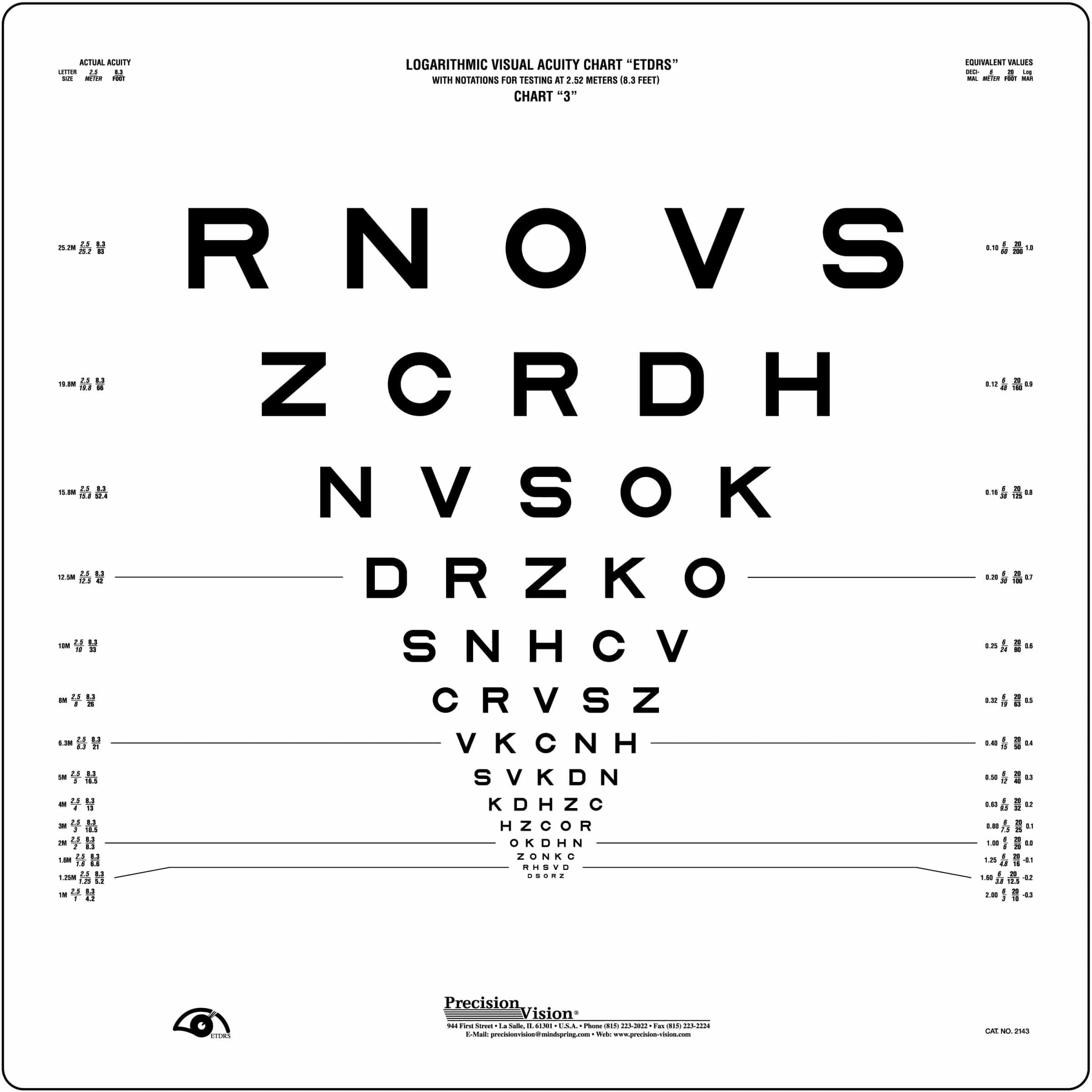
Sloan Letter Revised Series ETDRS Charts (2.5 Meter) Precision Vision
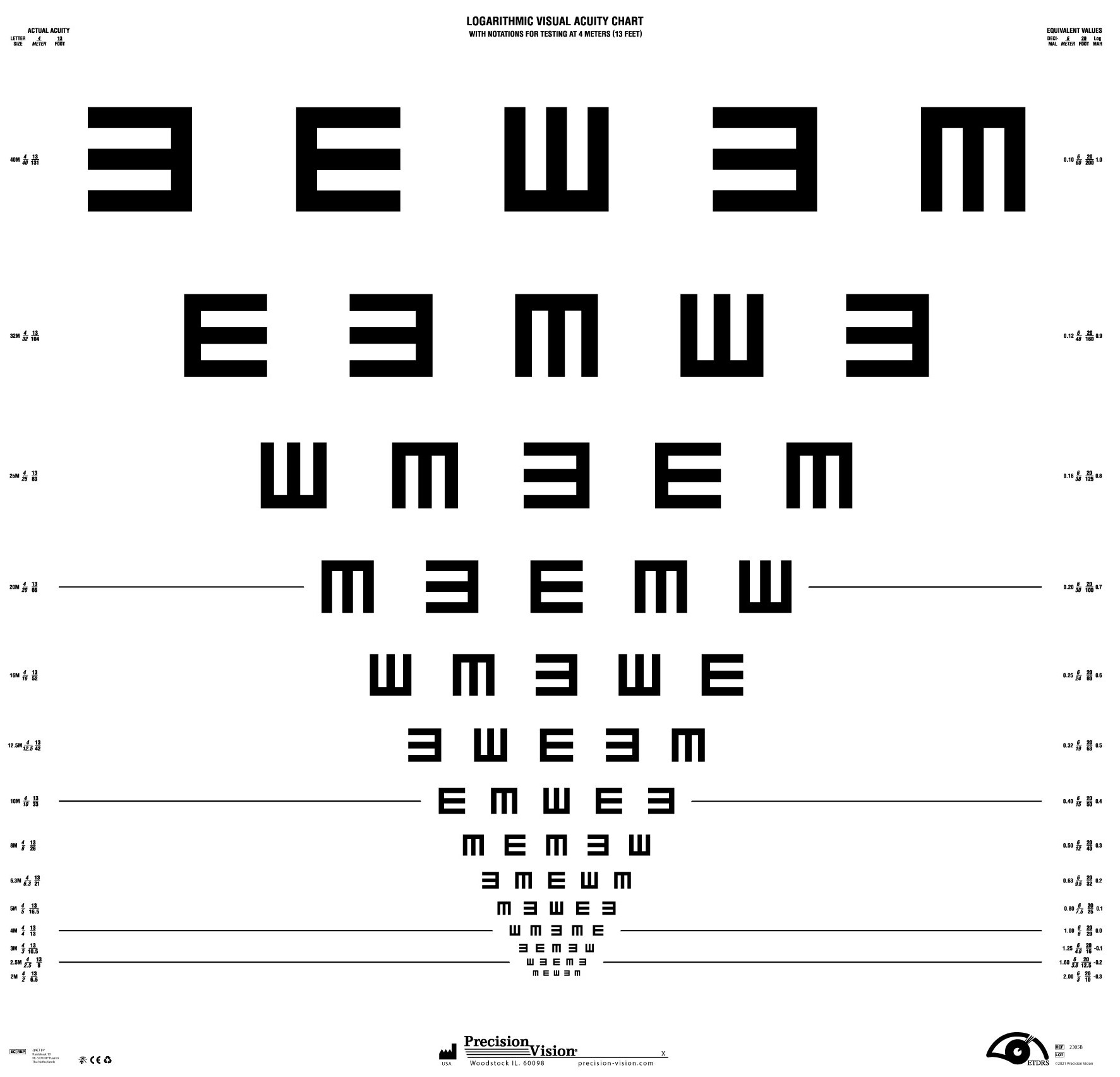
Tumbling E Series ETDRS® (Chart 3) Precision Vision
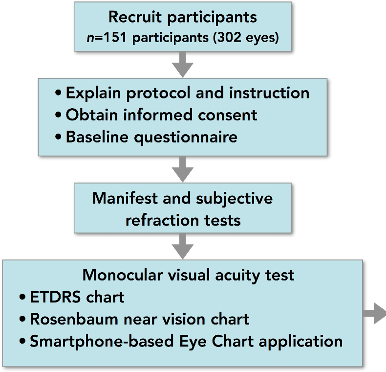
Figure 1 from Comparison of Visual Acuity Measurement Using Three Methods Standard ETDRS Chart

Original Series Sloan Letter ETDRS Chart R Jutron Vision

Comparison of visual acuity values on Snellen, ETDRS charts, decimal... Download Scientific
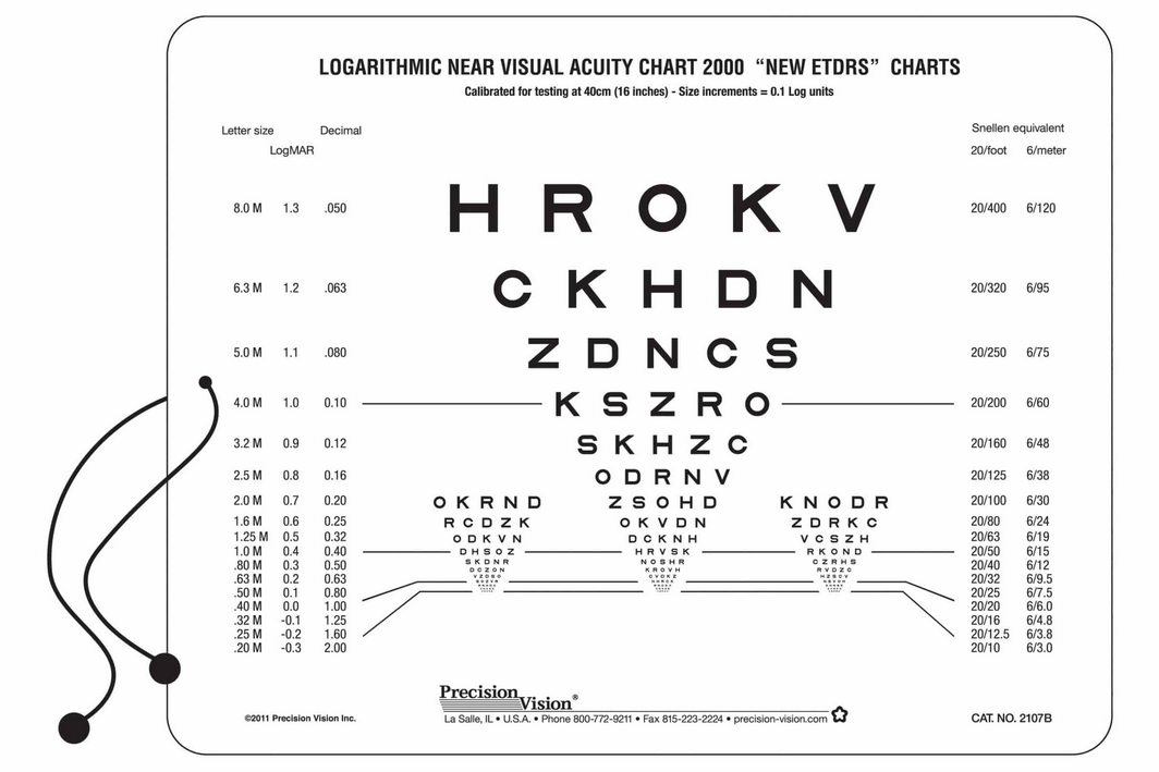
Near chart ETDRS letters 40cm, 2sided, scrambled "B" Near Tests Visual Acuity VISUS
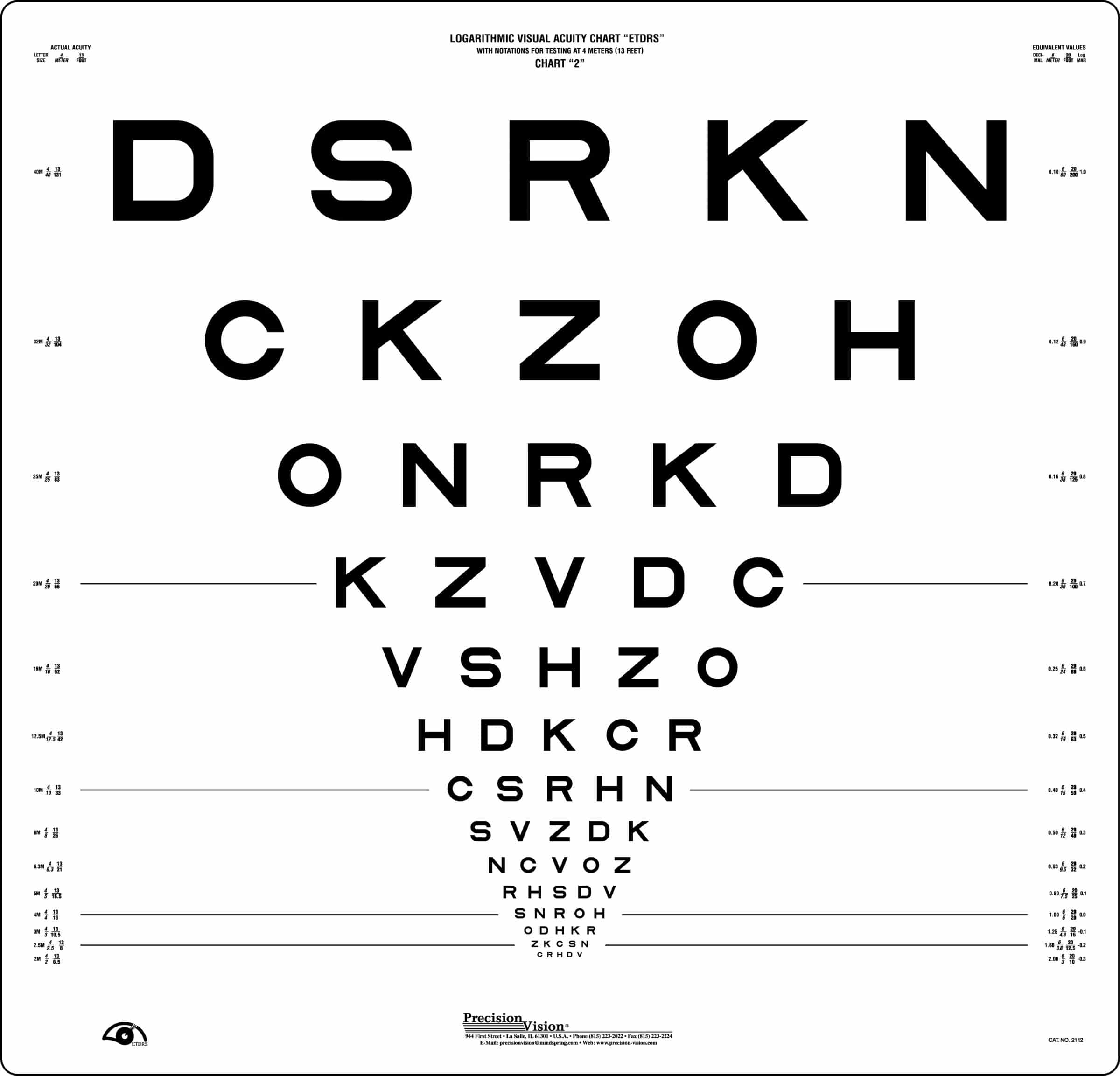
ETDRS Charts Precision Vision

ETDRS chart for measuring visual acuity. Download Scientific Diagram

Early Treatment Diabetic Retinopathy Study (ETDRS) chart. Download Scientific Diagram
ETDRS Charts Precision Vision
Csme Was Diagnosed Only By Clinical.
Classification Diabetic Retinopathy Falls Into Two Main Classes:
The Early Treatment Of Diabetic Retinopathy Study (Etdrs) Chart Uses A Logmar Design.
Related Post: